
A mother’s body undergoes several changes during the 9-months of pregnancy, particularly inside the womb. Soon after conception, the placenta and umbilical cord start to develop in the mother’s uterus. They play a significant role as the baby’s support system, by providing oxygen, food, and waste removal for the growing fetus.
The placenta is a temporary organ, and the umbilical cord is a tube-like lifeline that connects the fetus to the placenta. This physical link allows the fetus to move freely inside the amniotic sac, also known as the “bag of water”, which surrounds a fetus during pregnancy. The amniotic sac is a thin, tough, transparent pair of membranes that surround the amniotic fluid, placenta, and umbilical cord. As the fetus develops, the amniotic sac grows. The main function of the amniotic sac is to protect the fetus, as the amniotic fluid helps cushion the fetus from bumps and regulate its body temperature.
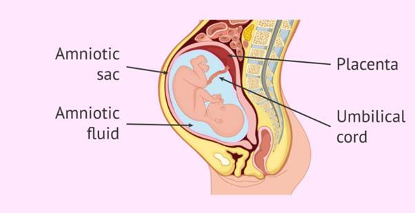
Development and structure of the placenta:
Development: According to the Cleveland Clinic, the placenta begins to form after a fertilized egg implants in the uterine lining and it has the same genetic makeup as the fetus. It forms at the spot where the fertilized egg implants on the inner lining of the uterus. It continues to grow during pregnancy to support the fetus, starting as a few cells and growing to end up several centimeters long. It starts to produce hormones by the end of the first trimester (12 weeks), which is also when first trimester symptoms such as nausea and fatigue usually go away.
Delivery of the placenta occurs shortly after the baby is born, usually within 30 minutes, and is called afterbirth or the third stage of labour. After a vaginal delivery, the uterus will continue to contract to get rid of the placenta. With a C-section, the placenta is removed by the medical practitioner through the same incision the baby came through.
Structure: The placenta has three main parts, namely the side that faces the fetus and where the umbilical cord is attached, then there’s the surface that’s attached to the uterine wall, and thirdly the cavity between these two surfaces. The placenta is deep red in colour, as it mostly contains blood vessels. It looks like a big disc of bumpy tissue and has a gel-like consistency. The average size of a mature placenta is almost 22 centimeters (cm) long and the center is about 2.5 cm thick.
The placenta appears to move, resulting from the uterus expanding as the pregnancy continues. Most placentas appear to “move” to the top or side of the uterus by 32 weeks of pregnancy, as the fetus grows.
Role of the placenta:
According to Thrombosis Research, the placenta is a highly specialized organ of pregnancy that supports the normal growth and development of the fetus. The growth and function of the placenta are precisely regulated and coordinated to ensure the exchange of oxygen, nutrients, and waste products such as carbon dioxide between the regulatory systems of the mother and the fetus. This exchange occurs through tiny blood vessels called villi, within which the fetal blood is separated by only three or four cell layers (called the placental membrane) from the maternal blood.
The placenta metabolizes a number of substances and can release metabolic products into fetal and/or maternal circulation. It can help to protect the fetus against infections and maternal diseases. Once the placenta starts to produce hormones at the end of the first trimester, these hormones help to maintain the pregnancy and help the fetus grow. Hormones such as estrogen and progesterone (amongst others) are released into both the maternal and fetal circulatory systems and affect functions such as metabolism, fetal growth, and the process of giving birth.
The placenta acts as a barrier that keeps the mother’s blood and the fetus’s blood separate, while letting vital substances pass through. Closer to the delivery date, the placenta passes antibodies from the mother’s immune system to the fetus to trigger immunity, which sticks with the baby for several months after birth.
Interestingly, the placenta is devoid of nerves. As a result, any communication between the placenta and the mother and/or fetus would normally occur through blood-borne substances.
Development and structure of the umbilical cord:
Development: Umbilical cord development begins during the embryonic period, around week 3, with the formation of the connecting stalk. By week 7, the umbilical cord has fully formed, composed of the connecting stalk, vitelline duct (connecting the yolk sac to the primitive midgut), and umbilical vessels. The umbilical vessels carry the fetal blood back and forth to the placenta.
Elongation of the umbilical cord occurs primarily in the second trimester. The average umbilical cord is 50 to 60 cm in length, 2 cm in diameter, with up to 40 helical spirals, which provide structural resilience.
Structure: Temporary embryonic structures regress near the end of the first trimester. A mature cord contains two umbilical arteries and one large umbilical vein. The blood vessels are embedded in a protective, gelatinous extracellular matrix known as Wharton’s jelly, which prevents compression and kinking of the blood vessels. The entire outer surface of the cord is covered by a tubular protective layer derived from the amniotic membrane, known as the amniotic sheath.
Role of the umbilical cord:
According to the I.D. Institute for Stem cell and Genome Research in Ukraine, the umbilical cord is the lifeline that connects the fetus to the placenta and plays a vital role in the growth and development of the fetus, such as:
- Nutrient and Oxygen Transport: The umbilical cord acts as a bridge between the mother and fetus, with the umbilical vein transporting oxygen- and nutrient-rich blood from the placenta to the fetus, ensuring it receives the necessary elements for its growth and development.
- Waste Removal: The two arteries in the umbilical cord transport waste and carbon dioxide from the fetus to the placenta, where they are removed via the mother’s blood.
- Protection: The umbilical cord protects the fetus from external shocks and stresses, due to its spirals and flexible structure, as well as the gelatinous substance (Wharton’s jelly) that surround the blood vessels.
- Source of Stem Cells: Umbilical cord blood contains a large amount of stem cells, which are unspecialized cells that can develop into various cell types in the body.
- Therapeutic Potential: Studies have shown that stem cells found in umbilical cord blood can be used to treat many diseases, for example blood diseases (such as leukemia and thalassemia); immune system diseases; some types of cancer; and spinal cord injuries. Stem cells found in umbilical cord blood are often more compatible with the patient’s body than stem cells taken from other sources, reducing the risk of rejection.
The umbilical cord is regarded as a natural treasure with tremendous therapeutic potential. With continued medical research and developments, the umbilical cord is expected to play an increasingly important role in treating diseases and improving human health. In some countries, the demand for cord blood banking is rising exponentially. Umbilical cord blood banking or stem cell preservation is a process of collecting umbilical cord blood, extracting its stem cells, and cryogenically freezing it for future medical use.
Conclusion:
The placenta and umbilical cord are highly specialized and essential structures that support the fetus throughout pregnancy. The placenta provides a vital interface between the mother and fetus, facilitating the exchange of oxygen, nutrients, hormones, and waste products while also contributing to immune protection and the maintenance of pregnancy. The umbilical cord, through its two arteries and one vein protected by Wharton’s jelly, provides the physical connection between the fetus and placenta and ensures the efficient transport of oxygen, nutrients, and waste products. Together with the amniotic sac and fluid, these structures create a protected environment in which the fetus can grow and develop.
Beyond their importance during fetal development, the placenta and umbilical cord also have significance after birth. The placenta is used in some circles for placenta encapsulation, while umbilical cord blood contains valuable stem cells that have established therapeutic applications in the treatment of certain blood and immune-system disorders.
Understanding the placenta and umbilical cord is important, not only for appreciating normal pregnancy and fetal development, but also for advancing healthcare and exploring future medical therapies. Ongoing research continues to investigate their wider potential.
References:
Placenta. Published 15 December 2025. Cleveland Clinic. USA. (A nonprofit multi-specialty medical centre that integrates clinical and hospital care with research and education.) (www.clevelandclinic.org)
Placenta: How it works, what’s normal. Published 8 March 2024. Mayo Clinic. USA. (www.mayoclinic.org)
Growth and function of the normal human placenta. Published in the journal Thrombosis Research. Volume 114, Issues 5-6, 2004. Science Direct. (www.sciencedirect.com)
The umbilical cord and its important role: An overview. Published 6 March 2026. I.D. Institute for Stem Cell and Genome Research. Ukraine. (www.idstemgenomeinstitute.com)
Placenta & umbilical cord. Published 25 September 2021. Biocell. A stem cell preservation bank in India. (www.biocell.in)
Amniotic sac. Published 11 September 2024. Cleveland Clinic. USA. (A nonprofit multi-specialty medical center that integrates clinical and hospital care with research and education.) (www.clevelandclinic.org)
Embryology, umbilical cord. Published 17 April 2024. StatPearls. National Centre for Biotechnology Information. US National Library for Medicine. National Institutes of Health. USA. (www.ncbi.nlm.nih.gov)
HEALTH INSIGHT